- Преподавателю
- Иностранные языки
- Дидактическое пособие по английскому языку для студентов медицинских колледжей Анатомия в картинках
Дидактическое пособие по английскому языку для студентов медицинских колледжей Анатомия в картинках
| Раздел | Иностранные языки |
| Класс | - |
| Тип | Другие методич. материалы |
| Автор | Лаврентьева И.А. |
| Дата | 26.10.2015 |
| Формат | doc |
| Изображения | Есть |

Орловский базовый медицинский колледж
Дидактическое пособие
по английскому языку
«LOOK & LEARN» / «Смотри и учись!»
(Анатомия в картинках)
Тексты для дополнительного чтения
для самостоятельной работы студентов
2 курса
по разделу «Мы изучаем анатомию».
Составила: Лаврентьева И.А.
Орел 2014 г.
Skeleton
Introduction
This system has 206 bones and associated cartilage, tendons, and ligaments. Because bone is rigid, it gives the body a framework, maintains its shape, and protects vital organs. Bones provide a place for muscles and supporting structures to attach, and, with the movable joints, form a system of levers upon which muscles can act to produce body movements. A joint is a place of union between two or more bones that may be movable or immovable. Bone also functions as a site for mineral storage and blood cell formation. Tendons and ligaments are strong bands of fibrous connective tissue that attach muscles to bones, and bones to bones, respectively.
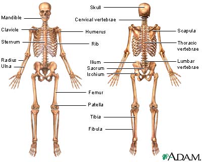

Axial skeleton
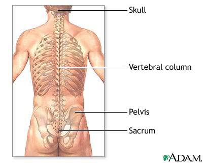
The skeleton has two parts: the axial skeleton and the appendicular skeleton.
The axial skeleton includes the skull, the hyoid bone, the vertebral column (spine, sacrum, and coccyx), the sternum, and the ribs. Its components are aligned along the long axis of the body.
The appendicular skeleton includes the bones of the upper extremities (arms, forearms, and hands), the pectoral (shoulder) girdle, the pelvic (hip) girdle, and the bones of the lower extremities (thigh, knee, leg, and foot). Its components are outside the body main axis.
Bone tissue stores and releases ionic minerals that affect homeostasis (stable internal environment) of the body. Chief among these minerals is calcium, which is necessary for proper functioning of the muscles and nervous system. Endocrine system hormones regulate calcium release and storage.
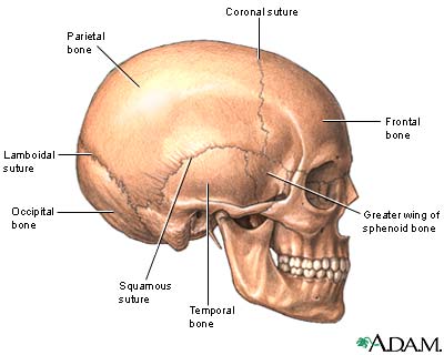
Cranial bones are flat, rounded, and fused to protect the brain. The eight cranial bones are the frontal (1), parietal (2), temporal (2), occipital (1), sphenoid (1), and ethmoid (1). Two cranial bones meet at a suture (immovable joint). The four major sutures are the coronal suture (between the parietal and frontal bones), the lambdoidal suture (between the occipital and parietal bones), the sagittal suture (between the two parietal bones), and the squamous suture (between the parietal and temporal bones).
Facial bones provide a framework for the facial muscles, form eye sockets, and form jaws for the teeth. The fourteen facial bones are the vomer (1), maxilla (2), zygomatic (2), palatine (2), lacrimal (2), nasal (2), inferior nasal conchae (2), and mandible (1). The hinged mandible (jaw bone) moves freely during mastication (chewing) and speech.
The vertebral column has three groups of vertebrae and two sets of fused bones. These vertebrae include seven cervical (neck) vertebrae, twelve thoracic (upper back) vertebrae, and five lumbar (lower back) vertebrae. Five fused vertebrae form the sacrum and from three to five fused small vertebrae form the coccyx (tail bone). The vertebrae form a column of bone that protects the spinal cord. The thoracic vertebrae have facets (indentations) upon their surfaces that articulate (meet) with the ribs.
The twelve pairs of ribs are long, flattened, and curved bones that form a protective cage for the heart, lungs, and other internal organs. The vertebrosternal (true) ribs are the first seven ribs; they are "true" because they attach directly to the sternum (breast bone). Ribs eight through twelve are the false ribs because they indirectly attach to the sternum or they lack a sternal attachment. Ribs eight through ten are the vertebrochondral ribs because they attach indirectly to the sternum by cartilage. Ribs eleven and twelve are called floating (vertebral) ribs because they do not attach to the sternum. Instead, their floating position allows them to bend sideways while providing protection for the kidneys.
Appendicular skeleton
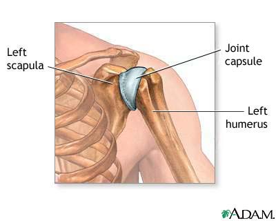
In the appendicular skeleton, the pectoral girdle bones include two scapulae (shoulder blades) and two clavicles (collar bones). The scapula is in the upper back and articulates with two bones: the humerus and the clavicle. Because the scapula is part of the shoulder joint, the scapula must be mobile to allow the upper extremities freedom of movement. The clavicle articulates with the sternum and the scapula, giving support to the pectoral girdle and adding stability to the shoulder joint.
The bones of the upper extremities are the humerus, ulna, radius, carpals (wrist bones), metacarpals (hand bones), and phalanges (finger bones). The humerus in the arm articulates with the scapula at the shoulder joint and with the ulna and radius at the elbow. The radius and ulna in the forearm articulate with the carpals at the wrist. The ulna articulates with the humerus and forms the elbow. The carpals are small, flat, irregularly shaped wrist bones. They articulate with the metacarpals in the hand. The metacarpals articulate with the phalanges.
In the pelvic girdle are two hip bones. Each coxa (hip bone) forms from fused bones. The two coxae, sacrum, and coccyx form the pelvis, a bowl-shaped cavity that supports and protects many abdominal organs. The three fused bones of each coxa are the ilium, ischium, and pubis.
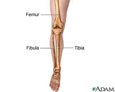
The pelvic girdle articulates with the femur (thigh bone) at the acetabulum (hip joint) and with the sacrum at the sacroiliac joint. Also, the coxae articulate with each other at the pubic symphysis, a joint with limited movement. The femur is the longest and heaviest bone in the body and helps in weight-bearing while standing.
At the knee, the femur articulates with the tibia (shin bone). Suspended within muscle tendons at the front of the knee joint is the patella (kneecap). The patella is an example of a sesamoid bone (small bone) that is within a tendon.
Attaching to the lateral (outer side) of the tibia is the fibula (leg bone). The fibula provides points of attachment for muscles of the foot and leg and increases the lateral stability of the ankle. The fibula is not a weight-bearing bone like the tibia.
In the foot are specialized bones designed for weight-bearing. Among these are the tarsals (ankle bones), metatarsals (foot bones), and phalanges (toe bones). These foot bones form a system of arches that allow the foot to support much weight.
Ossification and reconstruction
Ossification (bone growth) begins during fetal development. Before birth every skeleton bone appears as a fibrous membrane template (membranous bone) or a cartilaginous template (cartilaginous bone). These templates form the basic shapes that mature bone replaces. Membranous bone develops through intramembranous ossification. Cartilaginous bones develop through endochondral ossification.
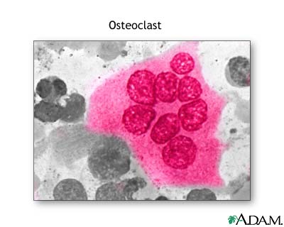
Despite its hardness and relative inflexibility, bone is one of the most changing tissues of the body. At the bone tissue cellular level, reorganization and replacement of the solid matrix occurs continuously. Two types of cells are primarily responsible for this reconstructive process: the osteoblasts and the osteoclasts.
Osteoblasts form new bone tissue in response to more demands on bone. When a person is physically active, the rate of osteoblast activity increases, which thickens and strengthens the bones. When muscles tense and flex, their tendons pull on the bones. This action causes the osteoblasts to secrete proteins that form lamellar (compact) bone and cancellous (spongy) bone with texture that resembles trabeculae (a web-like network of tunnels).
Within compact bone is a system of canals that contain blood vessels, nerves, and lymph vessels. These canals form functional units called osteons (Haversian systems). The osteon is a tube-like structure containing a central (Haversian) canal through which nerves and blood vessels pass. Surrounding the central canal is bone matrix, a substance produced by osteocytes. Within the osteon, osteocytes form lamellae, concentric layers of hard bone matrix. As osteoblasts create lamellae, these osteoblasts become trapped within the compact bone and are called osteocytes.
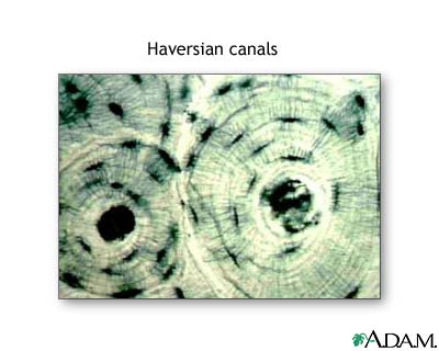
Osteocytes lie within lacunae, small pockets between the lamellae. Canaliculi, tiny canals, connect the lacunae. Canaliculi also connect with the central canal. These connections allow nutrients to pass through the canaliculi to the trapped osteocytes within the dense lamellae.
Osteoclasts work with osteoblasts. These osteoclasts dissolve bone tissue by secreting enzymes. Their actions help healthy individuals keep bones light and airy. They also regulate calcium and phosphate concentrations in body fluids. When osteoclast activity is not balanced by new bone formation, osteoporosis, a degenerative bone disease, occurs.
Bone marrow
Within the long bones are two types of bone marrow: red marrow and yellow marrow. The yellow marrow has fatty connective tissue and fills the marrow cavity. During starvation, the body uses the fat in yellow marrow for energy.
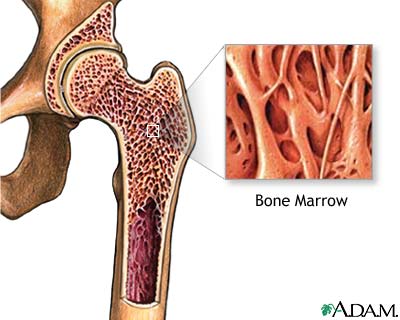
The red marrow of some bones is an important site for blood cell production. Here all erythrocytes (red blood cells), platelets, and most leukocytes (white blood cells) form in adults. From the red marrow, erythrocytes, platelets, and leukocytes migrate to the blood to do their special tasks. Red blood cells carry oxygen and nutrients to the body tissues. Platelets help in blood clotting. White blood cells help fight disease and infection.
Joints
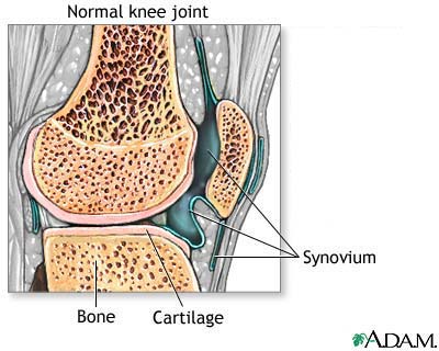
Bones of the skeleton articulate at joints. Joints form three categories of movability: freely movable, slightly movable, and immovable. Freely movable joints are also called synovial joints. A typical synovial joint has a joint capsule, a synovial membrane, synovial fluid, a joint cavity, and articular cartilage. A joint capsule surrounds the joint, supporting and stabilizing it. The synovial membrane is within the joint capsule. This membrane closely surrounds the joint and forms a joint cavity. The synovial membrane secretes synovial fluid that lubricates the articular surfaces of the joint. In some joints, the synovial membrane extends outside the joint capsule to form a bursa. The bursa cushions the joint. Bursae are in the knee, elbow, shoulder, and hip. Articular cartilage covers the articular surfaces of synovial joints to prevent excess wear and tear as they move against each other.
Six types of synovial joints are hinge, ball-and-socket, pivot, condyloid (angular or ellipsoidal), plane (gliding), and saddle. The elbow is an example of a hinge joint. Here, the convex and concave articulating bones allow movement along one plane, similar to a door.
The shoulder and hip are the only ball-and-socket joints in the body. In this type of joint, one bone has a spherical head that articulates with a corresponding concavity. This joint frees the joint to move in many directions. In a pivot joint, one round-shaped articulating bone fits within a corresponding depression on another bone. This joint allows one bone to rotate against the other. An example is the radioulnar joint (joint of the radius and ulna) in the forearm. In a condyloid (angular) joint, one bone has an oval articulating head that rests within an oval concavity. This joint permits angular movement of the bones. The metacarpophalangeal joint (junction between the metacarpals and phalanges) of the hand are examples of condyloid joints.
Plane joints have two flat bones joined. The sole movement of the bones is short gliding motions. An example of this joint is the intertarsal joint (junction between the tarsal bones) of the feet. Saddle joint bones have convex and concave surfaces similar to a saddle. This joint allows the joint to move in many directions. The carpometacarpal joint of the thumb is an example saddle joint.
As their name implies, amphiarthrosis joints (slightly movable joints) have limited movement. The two types of amphiarthrosis joints are syndesmosis (fibrous) and symphysis (cartilaginous). A syndesmosis joint occurs when two bones join by a section of cartilage. The junction between the tibia and fibula is an example. A symphysis joint forms when two bones fuse by a fibrocartilage pad. Typical symphysis joints are between the pubic symphysis (pubic bones in the pelvis), and in the vertebral column between individual vertebrae. Intervertebral discs act as weight-bearing shock absorbers for walking, jumping, and lifting.
An immovable joint is called a synarthrosis. The two types of this joint are sutures and gomphoses. Sutures are joined by short fibers of dense fibrous connective tissue and are in the skull. The single example of a gomphosis joint is the teeth sitting within their sockets. An example of a bony fusion joint is the fusion of the three bones forming a coxa (hip bone): the ilium, ischium, and pubis.
Ligaments link bones. These ligaments, sheets of strong, fibrous connective tissue, are identical with the muscular system tendons. The only difference is their function: tendons attach muscle to bone and ligaments attach bone to bone.
Nervous system
Introduction
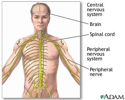
As the most complex system, the nervous system serves as the body control center and communications electrical-chemical wiring network. As a key homeostatic regulatory and coordinating system, it detects, interprets, and responds to changes in internal and external conditions. The nervous system integrates countless bits of information and generates appropriate reactions by sending electrochemical impulses through nerves to effector organs such as muscles and glands. The brain and spinal cord are the central nervous system (CNS); the connecting nerve processes to effectors and receptors serve as the peripheral nervous system (PNS). Special sense receptors provide for taste, smell, sight, hearing, and balance. Nerves carry all messages exchanged between the CNS and the rest of the body.
CNS: neurons, brain, spinal cord
The neuron transmits electric signals like an electric wire. The perikaryon (cell body) is the neuron central part. Dendrites, short branches, extend from the neuron. These input channels receive information from other neurons or sensory cells (cells that receive information from the environment). A long branch, the axon, extends from the neuron as its output channel. The neuron sends messages along the axon to other neurons or directly to muscles or glands.
Neurons must be linked to each other in order to transmit signals. The connection between two neurons is a synapse. When a nerve impulse (electrical signal) travels across a neuron to the synapse, it causes the release of neurotransmitters. These chemicals carry the nerve signal across the synapse to another neuron.
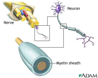
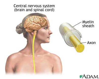
Nerve impulses are propagated (transmitted) along the entire length of an axon in a process called continuous conduction. To transmit nerve impulses faster, some axons are partially coated with myelin sheaths. These sheaths are composed of cell membranes from Schwann cells, a type of supporting cell outside the CNS. Nodes of Ranvier (short intervals of exposed axon) occur between myelin sheaths. Impulses moving along myelinated axons jump from node to node. This method of nerve impulse transmission is saltatory conduction.
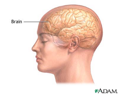
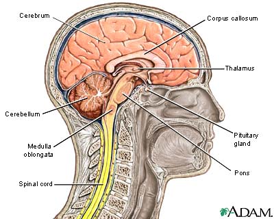
The brain has billions of neurons that receive, analyze, and store information about internal and external conditions. It is also the source of conscious and unconscious thoughts, moods, and emotions. Four major brain divisions govern its main functions: the cerebrum, the diencephalon, the cerebellum, and the brain stem.
The cerebrum is the large rounded area that divides into left and right hemispheres (halves) at a fissure (deep groove). The hemispheres communicate with each other through the corpus callosum (bundle of fibers between the hemispheres). Surprisingly, each hemisphere controls muscles and glands on the opposite side of the body. Comprising 85 percent of total brain weight, the cerebrum controls language, conscious thought, hearing, somatosensory functions (sense of touch), memory, personality development, and vision.
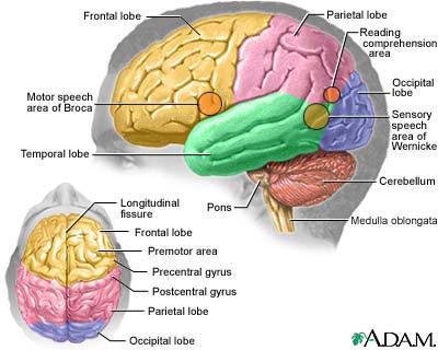
Gray matter (unmyelinated nerve cell bodies) composes the cerebral cortex (outer portion of the cerebrum). Beneath the cortex lies the white matter (myelinated axons). During embryonic development, the cortex folds upon itself to form gyri (folds) and sulci (shallow grooves) so that more gray matter can reside within the skull cavity.
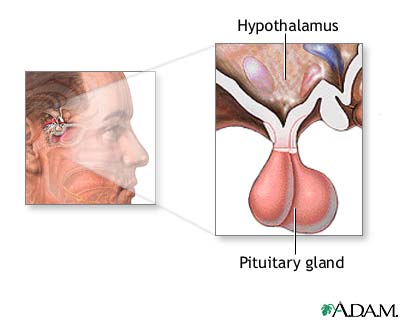
The diencephalon forms the central part of the brain. It consists of three bilaterally symmetrical structures: the hypothalamus, thalamus, and epithalamus. The hypothalamus 'master switchboard' resides in the brain stem upper end. It controls many body activities that affect homeostasis (maintenance of a stable internal environment in the body).
The hypothalamus is the main neural control center (brain part that controls endocrine glands). The pituitary gland lies just below the hypothalamus. The pituitary gland is a small endocrine gland that secretes a variety of hormones (organic chemicals that regulate the body's physiological processes). When the hypothalamus detects certain body changes, it releases regulating factors (chemicals that stimulate or inhibit the pituitary gland). The pituitary gland then releases or blocks various hormones. Because of this close association between the nervous and endocrine systems, together they are called the neuroendocrine system.
The hypothalamus also regulates visceral (organ-related) activities, food and fluid intake, sleep and wake patterns, sex drive, emotional states, and production of antidiuretic hormone (ADH) and oxytocin. The pituitary gland produces both these hormones.
The thalamus is a relay and preprocessing station for the many nerve impulses that pass through it. Impulses carrying similar messages are grouped in the thalamus, then relayed to the appropriate brain areas.
The epithalamus is the most dorsal (posterior) portion of the diencephalon. It contains a vascular network involved in cerebrospinal fluid production. Extending from the epithalamus posteriorly is the pineal body, or pineal gland. Its function is not yet fully understood; it is thought to control body rhythms.
At the rear of the brain is the cerebellum. The cerebellum is similar to the cerebrum: each has hemispheres that control the opposite side of the body and are covered by gray matter and surface folds. In the cerebellum, the folds are called folia; in the cerebrum, sulci. The vermis (central constricted area) connects the hemispheres. The cerebellum controls balance, posture, and coordination.
The brain stem connects the cerebrum and cerebellum to the spinal cord. Its superior portion, the midbrain, is the center for visual and auditory reflexes; examples of these include blinking and adjusting the ear to sound volume. The middle section, the pons, bridges the cerebellum hemispheres and higher brain centers with the spinal cord. Below the pons lies the medulla oblongata; it contains the control centers for swallowing, breathing, digestion, and heartbeat.
The reticular formation extends throughout the midbrain. This network of nerves has widespread connections in the brain and is essential for consciousness, awareness, and sleep. It also filters sensory input, which allows a person to ignore repetitive noises such as traffic, yet awaken instantly to a baby's cry.
The spinal cord is a continuation of the brain stem. It is long, cylindrical, and passes through a tunnel in the vertebrae called the vertebral canal. The spinal cord has many spinal segments, which are spinal cord regions from which pairs (one per segment) of spinal nerves arise. Like the cerebrum and cerebellum, the spinal cord has gray and white matter, although here the white matter is on the outside. The spinal cord carries messages between the CNS and the rest of the body, and mediates numerous spinal reflexes such as the knee-jerk reflex.
Meninges, three connective tissue layers, protect the brain and spinal cord. The outermost dura layer forms partitions in the skull that prevents excessive brain movement. The arachnoid middle layer forms a loose covering beneath the dura. The innermost pia layer clings to the brain and spinal cord; it contains many tiny blood vessels that supply these organs.
Another protective substance, cerebrospinal fluid, surrounds the brain and spinal cord. The brain floats within the cerebrospinal fluid, which prevents against crushing under its own weight and cushions against shocks from walking, jumping, and running.
PNS: somatic (voluntary) nervous system, autonomic (involuntary) nervous system
The peripheral nervous system includes sensory receptors, sensory neurons, and motor neurons. Sensory receptors are activated by a stimulus (change in the internal or external environment). The stimulus is converted to an electronic signal and transmitted to a sensory neuron. Sensory neurons connect sensory receptors to the CNS. The CNS processes the signal, and transmits a message back to an effector organ (an organ that responds to a nerve impulse from the CNS) through a motor neuron.
The PNS has two parts: the somatic nervous system and the autonomic nervous system. The somatic nervous system, or voluntary nervous system, enables humans to react consciously to environmental changes. It includes 31 pairs of spinal nerves and 12 pairs of cranial nerves. This system controls movements of skeletal (voluntary) muscles.
Thirty-one pairs of spinal nerves emerge from various segments of the spinal cord. Each spinal nerve has a dorsal root and a ventral root. The dorsal root contains afferent (sensory) fibers that transmit information to the spinal cord from the sensory receptors. The ventral root contains efferent (motor) fibers that carry messages from the spinal cord to the effectors. Cell bodies of the efferent fibers reside in the spinal cord gray matter. These roots become nerves that innervate (transmit nerve impulses to) muscles and organs throughout the body.
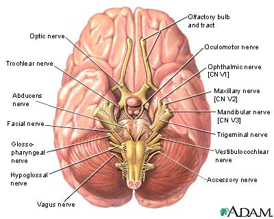
Twelve pairs of cranial nerves transmit from special sensory receptors information on the senses of balance, smell, sight, taste, and hearing. Cranial nerves also carry information from general sensory receptors in the body, mostly from the head region. This information is processed in the CNS; the resulting orders travel back through the cranial nerves to the skeletal muscles that control movements in the face and throat, such as for smiling and swallowing. In addition, some cranial nerves contain somatic and autonomic motor fibers.
The involuntary nervous system (autonomic nervous system) maintains homeostasis. As its name implies, this system works automatically and without voluntary input. Its parts include receptors within viscera (internal organs), the afferent nerves that relay the information to the CNS, and the efferent nerves that relay the action back to the effectors. The effectors in this system are smooth muscle, cardiac muscle and glands, all structures that function without conscious control. An example of autonomic control is movement of food through the digestive tract during sleep.
The efferent portion of the autonomic system is divided into sympathetic and parasympathetic systems. The sympathetic nerves mobilize energy for the 'Fight or Flight' reaction during stress, causing increased blood pressure, breathing rate, and bloodflow to muscles. Conversely, the parasympathetic nerves have a calming effect; they slow the heartbeat and breathing rate, and promote digestion and elimination. This example of intimate interaction with the endocrine system is one of many that explain why the two systems are called the neuroendocrine system.
The relationship between sensory and motor neurons can be seen in a reflex (rapid motor response to a stimulus). Reflexes are quick because they involve few neurons. Reflexes are either somatic (resulting in contraction of skeletal muscle) or autonomic (activation of smooth and cardiac muscle). All reflex arcs have five basic elements: a receptor, sensory neuron, integration center (CNS), motor neuron, and effector.
Spinal reflexes are somatic reflexes mediated by the spinal cord. These can involve higher brain centers. In a spinal reflex, the message is simultaneously sent to the spinal cord and brain. The reflex triggers the response without waiting for brain analysis. If a finger touches something hot, the finger jerks away from the danger. The burning sensation becomes an impulse in the sensory neurons. These neurons synapse in the spinal cord with motor neurons that cause the burned finger to pull away. This spinal reflex is a flexor, or withdrawal reflex.
The stretch reflex occurs when a muscle or its tendon is struck. The jolt causes the muscle to contract and inhibits antagonist muscle contraction. A familiar example is the patellar reflex, or knee-jerk reflex, that occurs when the patellar tendon is struck. The impulse travels via afferent neurons to the spinal cord where the message is interpreted. Two messages are sent back, one causing the quadriceps muscles to contract and the other inhibiting the antagonist hamstring muscles from contracting. The contraction of the quadriceps and inhibition of hamstrings cause the lower leg to kick, or knee-jerk.
Sense organs
The sense organs are highly specialized structures that receive information from the environment. These organs contain special sense receptors ranging from complex structures, such as eyes and ears, to small localized clusters of receptors, such as taste buds and olfactory epithelium (receptors for smell).
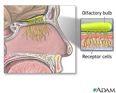
Smell and taste are chemical senses, which contain chemoreceptors that respond to chemicals in solution. Food chemicals dissolved in saliva stimulate taste receptors in taste buds. The nasal membranes produce fluids that dissolve chemicals in air. These chemicals stimulate smell receptors in olfactory epithelium. The chemical senses complement each other and respond to many of the same stimuli.
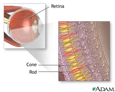
Photoreceptors, which include rods and cones, in back of the eye respond to light energy. Rods provide dim-light, black-and-white vision, and are the source of peripheral vision. Cones operate in bright light and provide color vision. Cones are most concentrated at the back center of each eye. Rods are more numerous than cones, and surround the cones. Information from the rods and cones travels via the optic nerve into the brain for interpretation.
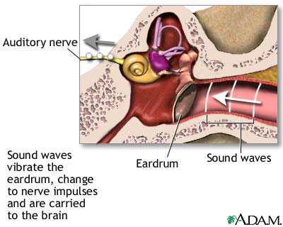
The ear has two specialized functions: sound wave detection and interpretation of the head position in space. Sound waves enter the outer ear through the external auditory canal (ear canal) and strike the tympanic membrane (eardrum). Vibration of the eardrum moves three ossicles (small bones) inside the middle ear, which in turn stimulate the organ of Corti (hearing receptor in the inner ear). Impulses travel from the organ of Corti through the vestibulocochlear nerve to be interpreted by the brain.
The ear also contains equilibrium (sense of balance) receptors. The vestibular apparatus, a group of equilibrium receptors in the inner ear, sense movement in space. Maculae receptors in the vestibule monitor static equilibrium (head position with respect to gravity when the body is still). Cristae receptors in the semicircular canals monitor dynamic equilibrium (movement). Impulses from the vestibular apparatus travel along the vestibulocochlear nerve to appropriate brain areas. These centers start responses that fix the eyes on objects and stimulate muscles to maintain balance.
Mechanoreceptors respond to mechanical energy forces: touch, pressure, stretching, and movement. Ranging in complexity from free nerve endings beneath the skin to more complex tactile receptors at the bases of hair, mechanoreceptors change shape when pushed or pulled.
Different types of skin receptors sense different environmental stimuli. Free nerve endings sense pain. Specialized receptors such as Merkel's discs and Meissner's corpuscles sense touch. Pacinian corpuscles sense deep pressure. Naked nerve endings are thought to be responsible for sensing temperature.
Other types of sensory receptors provide the brain information on the body. Interoreceptors in body organs inform the CNS about internal conditions such as hunger and pain. Proprioceptors in joints, tendons, and muscles detect changes in position of skeletal muscles and bones. This information allows humans to be aware the positions of their trunk and limbs without having to see them
Digestive system
Introduction
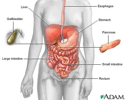
Nutrition permits us to take in and use food substances that the body converts to energy and body structure. The digestive system includes all the organs and glands involved in this process of eating and digesting. Starting in the mouth, a long muscular tube provides continual fluid and vital nutrients. The coiled intestines alone are about 24 feet long. After we consume food, the body mechanically and chemically breaks it down, then transports it for absorption and defecation (final waste removal). The digestive glands (salivary glands, pancreas, liver, and gallbladder) produce or store secretions that the body carries to the digestive tract in ducts and breaks down chemically.
Ingestion
Food processing begins with ingestion (eating). The teeth aid in mechanical digestion by masticating (chewing) food. Mastication permits easier deglutition (swallowing) and faster chemical breakdown in the digestive tract. During mastication, salivary glands secrete saliva to soften the food into a bolus (semi-solid lump). Saliva contains the salivary amylase enzyme, which digests carbohydrates (starches), and mucus (a thick liquid), which softens food into a bolus. Ingestion starts both chemical and mechanical digestion.
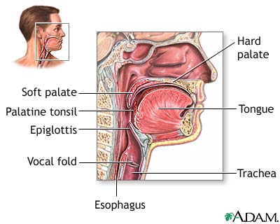
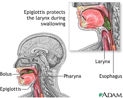
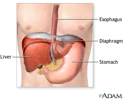
In deglutition, the tongue pushes the bolus toward the pharynx (throat) and into the esophagus, a muscular tube that leads from the throat to the stomach. To prevent food or liquid from entering the trachea (windpipe), the epiglottis (a small flap of tissue) closes over the opening of the larynx (voice box) during deglutition.
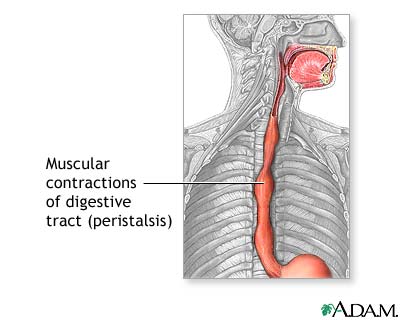
Upon entering the esophagus, peristalsis (wave-like contractions) of smooth muscle carries the bolus toward the stomach. Two layers of smooth muscle, the outer longitudinal (lengthwise) and inner circular, contract rhythmically to squeeze food through the esophagus. Throughout the digestive tract, smooth muscle peristalsis aids in transporting food.
From the esophagus, the bolus passes through a sphincter (muscular ring) into the stomach. All sphincters located in the digestive tract help move the digested material in one direction. When the stomach is empty, the walls are folded into rugae (stomach folds), which allow the stomach to expand as more food fills it.
Digestion: stomach
In the stomach, food undergoes chemical and mechanical digestion. Here, peristaltic contractions (mechanical digestion) churn the bolus, which mixes with strong digestive juices that the stomach lining cells secrete (chemical digestion). The stomach walls contain three layers of smooth muscle arranged in longitudinal, circular, and oblique (diagonal) rows. These muscles allow the stomach to squeeze and churn the food during mechanical digestion.
Powerful hydrochloric acid in the stomach helps break down the bolus into a liquid called chyme. A thick mucus layer that lines the stomach walls prevents the stomach from digesting itself. When mucus is limited, an ulcer (erosion of tissue) may form.
Food is digested in the stomach for several hours. During this time, a stomach enzyme called pepsin breaks down most of the protein in the food. Next, the chyme is slowly transported from the pylorus (end portion of the stomach) through a sphincter and into the small intestine where further digestion and nutrient absorption occurs.
Digestion and absorption: small intestine
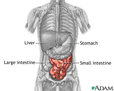
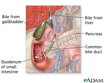
The small intestine is about 20 feet (6 meters) long and has three parts: the duodenum, jejunum, and ileum. The duodenum is where most chemical digestion takes place. Here, bile from the gallbladder and enzymes from the pancreas and intestinal walls combine with the chyme to begin the final part of digestion.
Bile liquid is created in the liver and stored in the gallbladder. Bile emulsifies (breaks into small particles) lipids (fats), which aids in the mechanical digestion of fats. The pancreas and gland cells of the small intestine secrete digestive enzymes that chemically break down complex food molecules into simpler ones. These enzymes include trypsin (for protein digestion), amylase (for carbohydrate digestion), and lipase (for lipid digestion). When food passes through the duodenum, digestion is complete.
From the duodenum, chyme passes to the jejunum and ileum. Here, tiny villi (finger-like projections) cover the walls of the small intestine. The cells that line the villi are covered with small projections called microvilli (brush border). These projections increase the surface area of the small intestine, allowing the chyme to contact more of the small intestine wall. The increased contact causes more efficient food absorption.
During food absorption, food molecules enter the bloodstream through the intestinal walls. Capillaries (microscopic blood vessels) within the villi absorb products of protein and carbohydrate digestion. Lymph vessels (lacteals) within the villi absorb products of fat digestion and eventually lead to the bloodstream.
From the small intestine, digested products travel to the liver, one of the body's most versatile organs. Hepatocytes (liver cells) detoxify (filter) blood of harmful substances such as alcohol and ammonia. And, hepatocytes store fat-soluble vitamins and excess substances such as glucose (sugar) for release when the body requires extra energy.
Absorption: large intestine
Once food has passed through the small intestine, it is mostly undigestible material and water. It enters the colon (large intestine), named for its wide diameter. The large intestine has six parts: the cecum, ascending colon, transverse colon, descending colon, sigmoid colon, and rectum.
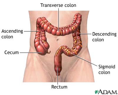
The large pouch-shaped cecum marks the beginning of the colon. Attached near the cecum bottom is the vermiform (worm-like) appendix. The appendix contains lymphoid tissue and intercepts pathogenic microorganisms that enter the digestive tract. Sometimes, fecal matter may become trapped in the appendix, resulting in appendicitis (infection and inflammation).
The other parts of the colon absorb water and minerals from the undigested food and compact the remaining material into feces. Defecation is the digestive process final stage: feces (undigested waste products) are carried to the rectum through peristalsis and eliminated through the anus.
Cardiovascular system
Introduction
Good Cardiovascular health is important in maintaining overall health and wellness. Cardiovascular Health is a new section, which will teach you how your heart and cardiovascular system work when healthy, and what happens when diseased. We will also explore disease prevention, therapeutic drug treatment, minimally invasive surgical procedures and open surgical procedures for treating diseases, which affect the heart and cardiovascular system.
The main role of the cardiovascular system in the body is to transport oxygen to all tissues in the body and for removing, from these same tissues, metabolic waste products. The system itself consists of the blood, the medium for exchanging oxygen, nutrients and waste products throughout the body, the blood vessels, the pipes through which the blood flows and the heart, the pump which forces blood to flow through the blood vessels. First, Lets take a closer look at the components of blood.
Components of Blood
Health conscious individuals should be especially concerned with maintaining a healthy cardiovascular system. While the heart, which is a powerful automatic pump, is the cardiovascular component we think of most often, the blood and the blood vessels are also vital to maintaining good health.
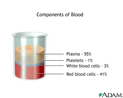
The average adult has between 5 to 6 liters of blood or blood volume. The blood carries oxygen and essential nutrients to all of the living cells in the body, and also carries waste products to systems that eliminate them. Most of the blood is made up of a watery, protein-laden fluid called plasma. A little less than half of this blood volume is composed of red and white blood cells, and other solid elements called platelets.
Platelets are responsible for coagulation of blood at the point of an injury to a blood vessel.
Without platelets, our blood would not be able to clot and hemorrhaging or uncontrolled bleeding would result. Hemophilia is a genetic condition, which results in individuals with no ability to clot. Also called bleeders, these individuals must periodically administer a clotting factor to their blood to prevent the constant bleeding, which occurs.
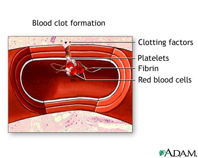
How Blood Clots
Let's examine how platelets work to form clots. Here's a cut section of a small artery. The traffic going by includes red blood cells carrying oxygen; platelets, which come from white blood cell fragments; and clotting factors, which help the blood to clot. When a blood vessel becomes damaged, as shown here, the blood cells and plasma begin oozing out into the surrounding tissue. This begins the clotting process. Platelets immediately begin to adhere to the cut edges of the artery; they release chemicals to attract even more platelets. Eventually a platelet plug is formed, and the external bleeding stops.
Inside, the clotting factors take a much more active role by creating a cascade of clotting activity. The clotting factors cause strands of blood-borne material, called fibrin, to stick together and seal the inside of the wound. Eventually, the cut blood vessel heals, and the blood clot dissolves after several days.
While platelets play an important role in clotting, red blood cells carry on the important job of carrying oxygen and other nutrients to all the tissues of the body and carrying waste products to the organs, which remove them from the body.
How Red Blood Cells Carry Oxygen
Red blood cells are the oxygen carriers. As they travel away from the heart, they traverse smaller and smaller arteries, finally arriving at the collections of microscopic blood vessels known as capillaries. Here, they exchange nutrients and oxygen for cellular waste products. The waste products are eventually eliminated from the blood stream through the urinary and respiratory systems.
The exchange of oxygen and nutrients between the red blood cells and the surrounding tissues occurs through a process called diffusion. In diffusion, when capillaries contain a high concentration of oxygen and nutrients, while the surrounding tissues contain a lower concentration, Oxygen and nutrients leave the capillaries and enter the tissues.
Conversely, when body tissues contain high concentrations of carbon dioxide and metabolic waste, while the capillaries contain a lower concentration, the waste products diffuse from the tissues into the capillaries and from there are carried by the venous system back toward the heart.
Blood Pressure
The red blood cells transport oxygen and waste products by flowing through the blood vessels. What causes blood to flow through the vessels is blood pressure. Just as water flows through pipes from areas of greater pressure to lesser, so too the blood flows through the body from areas of higher pressure to areas of lower pressure. Blood pressure is measured both as the heart contracts, which is called systole, and as it relaxes, which is called diastole. A systolic blood pressure of 120 millimeters of mercury is considered right in the middle of the range of normal blood pressures, as is a diastolic pressure of eighty. In common terms, this normal measurement would be stated as "120 over 80".
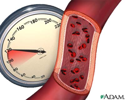
Normal blood pressure is important for proper blood flow to the body's organs and tissues. Each heartbeat forces blood to the rest of the body. The force of the blood on the walls of the arteries is called blood pressure. Blood pressure moves from high pressure near the heart to low pressure away from the heart. Blood pressure depends on many factors, including the amount of blood pumped by the heart. The diameter of the arteries through which blood is pumped is also an important factor. Generally, blood pressure is higher when more blood is pumped by the heart, and the diameter of an artery is narrow.
Systolic pressure is measured when the heart ventricles contract. Diastolic pressure is measured when the heart ventricles relax. Stressful situations can result in a temporary increase in blood pressure. If an individual were to have a consistent blood pressure reading of 140 over 90, he would be evaluated for having high blood pressure. If left untreated, high blood pressure can damage important organs, such as the brain and kidneys as well as lead to a stroke.
The Heart (The Pump)
The pressure and flow of the blood arise from the beating heart muscle, which is the pump of the cardiovascular system. To understand how the heart works lets take a brief look at the anatomy of the beating heart.
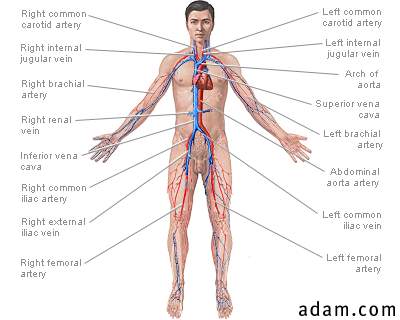
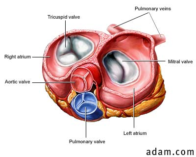
The heart is a four-chambered organ with four main vessels, which either bring blood to or carry blood away from the heart. The four chambers of the heart are: the right atrium, the right ventricle, the left atrium, and the left ventricle.
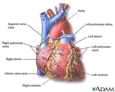
The great vessels that you see include the superior and inferior vena cava, which bring blood from the body to the right atrium. The pulmonary artery, which transports blood from the right ventricle to the lungs, and the aorta, the body's largest artery, which transports oxygen-rich blood from the left ventricle to the rest of the body.
If you look carefully, you can see a series of one-way valves that keep the blood flowing in one direction. The blood first enters the heart into the right atrium. A contraction of the right atrium then forces blood through the tricuspid valve and into the right ventricle. When the right ventricle contracts, the muscular force pushes blood through the pulmonary semilunar valve into the pulmonary artery.
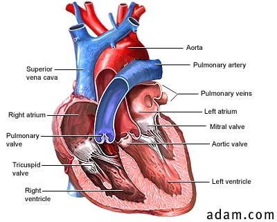
The blood then travels to the lungs, where it receives oxygen. Next, it drains out of the lungs via the pulmonary veins, and travels to the left atrium. From the left atrium, the blood is forced through the bicuspid valve into the critically important left ventricle. The left ventricle is the major muscular pump that sends the blood out to the body systems. When the left ventricle contracts, it forces the blood through the aortic semilunar valves and into the aorta. From here, the aorta and its branches carry blood to all the tissues of the body.
Respiratory system
Introduction
All the body cells metabolically consume oxygen, and discharge carbon dioxide. To cover this need, respiration takes place internally (at the cellular level) and externally (ventilation/breathing). Ventilation involves the inhalation of atmospheric air into the lungs via the nose and mouth through branching passageways, and the exhalation of carbon dioxide. The lung key function is to bring air and blood into intimate contact in the alveolar air sacs so that oxygen can enter the blood, and carbon dioxide can leave. At rest, humans breathe about twelve times a minute, bringing in approximately a pint of air. Exercise and certain diseases result in a marked increase of breathing. The respiratory system also is vital in maintaining normal blood pH and body temperature.
Lungs and air passages
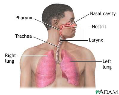
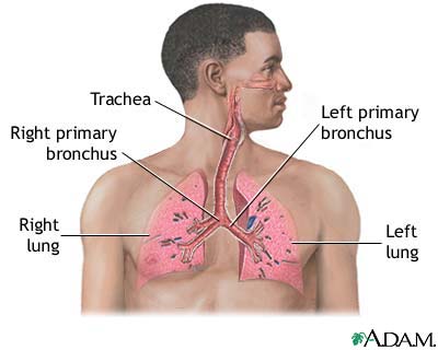
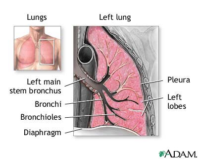
The lungs are paired organs that lie on either side of the heart and fill up the thoracic (chest) cavity. Inferior to (below) the lungs is the diaphragm, a broad thin muscle that separates the thoracic cavity from the abdominal (gut) cavity. On the medial (inner) surface of each lung is the hilus, where blood vessels, nerves, and bronchi (air passages) enter the lungs.
The lungs differ in size and shape. Because the heart is slightly larger on the left side, the left lung has a cardiac notch (indented border). The left lung is also slightly smaller than the right. Each lung is divided into lobes (partitions) by fissures. The right lung has three lobes: lower, middle, and upper. These horizontal and oblique fissures create these lobes. The left lung has upper and lower lobes that are divided by the oblique fissure.
Air enters the body through the mouth or nose. In the nose, thick hairs lining the nostrils prevent small objects from entering the nasal cavity. This cavity is lined with cells that produce mucus. Small foreign matter that enters the nasal cavities is trapped in the mucus, while tiny cilia (small hair-like projections) push the mucus to the pharynx (throat), where it is swallowed and digested in the stomach or expectorated.
From the pharynx, the air passes to the larynx, which is called the voice box because it contains the vocal cords. To prevent food or liquid from entering the larynx, the epiglottis (a small flap of tissue) closes over the opening of the larynx during deglutition (swallowing). If this process works improperly, a cough reflex expels the foreign material.
When air travels past the larynx, it enters the trachea (windpipe). The trachea is a strong tube containing rings of cartilage that prevents it from collapsing. The mucosa that lines the airway warms and moistens the air before it reaches the trachea.
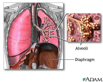
Within the lungs, the trachea branches into a left and right bronchus, which divide into increasingly smaller branches called bronchioles. The smallest bronchioles end in a cluster of air sacs, collectively called an acinus. The acinus comprises individual air sacs called alveoli. Alveoli are like small balloons that inflate and deflate with air during respiration.
Gas exchange
Gas exchange occurs in the lungs between the alveoli and a capillary network within the alveolar wall. Capillaries are microscopic blood vessels that exchange material between the blood and body tissues. In the lung capillaries, blood from tissues where cellular metabolism is occurring is called deoxygenated blood because it contains many carbon dioxide molecules and few oxygen molecules.
Respiration
The respiration process has two parts: inspiration (inhaling) and expiration (exhaling). During inspiration, the diaphragm contracts, moves downward, and causes the thoracic cavity volume to increase. Because the lungs are closely associated with the interior chest wall, they expand as the thoracic cavity expands. When the diaphragm relaxes (upward position), the thoracic volume decreases and the lungs partially deflate. This process is called expiration. The elastic recoil of the expanded thoracic wall and lungs also helps expiration.
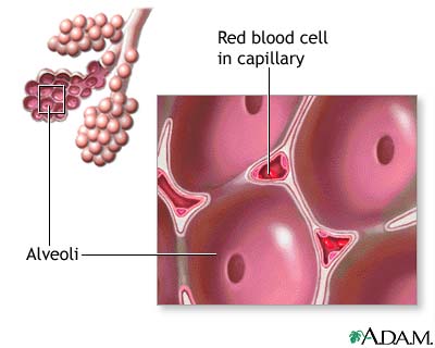
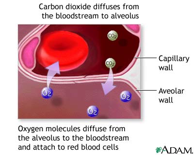
After inhalation, the alveoli contain many oxygen molecules. The alveoli are in close contact with the capillary network. This proximity enables the minuscule oxygen molecules to diffuse (pass freely) from the alveolus to the bloodstream, flowing from a region of higher concentration to a region of lower concentration. In the bloodstream, the oxygen attaches to red blood cells and is transported to the rest of the body. Likewise, carbon dioxide diffuses from the bloodstream into the alveolus where it is transported out of the body during exhalation.
During respiration, the pleurae (pleural membranes) help the lungs to expand and contract. These membranes are sacs that tightly cover the lungs and the chest inside wall. Between these two linings is a space called the pleural cavity that contains a thin layer of fluid. This fluid allows the lungs to move freely against the thoracic cavity inside.
Muscular system
Introduction
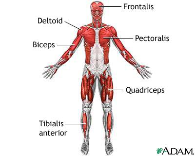
Over 600 skeletal muscles function for body movement through contraction and relaxation of voluntary, striated muscle fibers. These muscles are attached to bones, and are typically under conscious control for locomotion, facial expressions, posture, and other body movements. Muscles account for approximately 40 percent of body weight. The metabolism that occurs in this large mass-produces heat essential for the maintenance of body temperature.
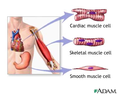
Cardiac muscle
Cardiac muscle is only in the heart and makes up the atria and ventricles (heart walls). Like skeletal muscle, cardiac muscle contains striated fibers. Cardiac muscle is called involuntary muscle because conscious thought does not control its contractions. Specialized cardiac muscle cells maintain a consistent heart rate.
Smooth muscle
Smooth muscle is throughout the body, including in visceral (internal) organs, blood vessels, and glands. Like cardiac muscle, smooth muscle is involuntary. Unlike skeletal and cardiac muscle, smooth muscle is nonstriated (not banded). Smooth muscle, which is extensively within the walls of digestive tract organs, causes peristalsis (wave-like contractions) that aids in food digestion and transport.
Except the heart, any action that the body performs without conscious thought is done by smooth muscle contractions. This includes diverse activities such as constricting (closing) the bronchioles (air passages) of the lungs or pupils of the eye or causing goosebumps in cold conditions.
Skeletal muscle
A skeletal muscle has regular, ordered groups of fascicles, muscle fibers, myofibrils, and myofilaments. Epimysium (thick connective tissue) binds groups of fascicles together. A fascicle has muscle fibers; perimysium (connective tissue) envelops the fascicle. Endomysium (connective tissue) surrounds the muscle fibers.
A muscle fiber divides into even smaller parts. Within each fiber are strands of myofibrils. These long cylindrical structures appear striped due to strands of tiny myofilaments. Myofilaments have two types of protein: actin (thin myofilaments) and myosin (thick myofilaments).
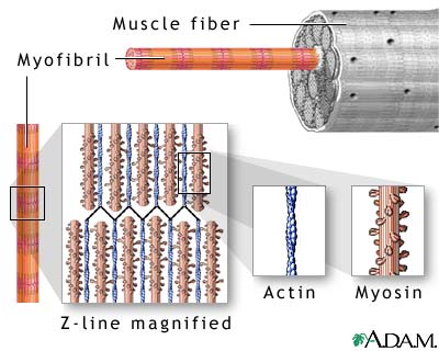
The actin and myosin myofilaments align evenly, producing dark and light bands on the myofibril. Each dark band depicts an area where the myofilaments overlap, causing the striated appearance of skeletal muscle.
All dark and light bands of the myofilaments have names. At the Z-line, actin strands interweave. The region between two Z-lines is a sarcomere, the functional unit of skeletal muscle. Muscle contraction occurs when overlapping actin and myosin myofilaments overlap further and shorten the muscle cell. The myofilaments keep their length. The overlapping of myofilaments is the basis for the sliding filament theory of contraction.
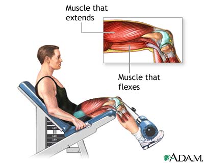
Skeletal muscle is a system of pairs that relax and contract to move a joint. For example, when front leg muscles contract, the knee extends (straightens) while back leg muscles relax. Conversely, to flex (bend) the knee, back leg muscles contract while front leg muscles relax. Some muscles are named for their ability to extend or flex a joint; for example, extensor carpiradialis longus muscle and flexor digitorum brevis muscle.
Tendons attach most skeletal muscles to bones. Tendons are strong sheets of connective tissue that are identical with ligaments. Tendons and ligaments differ in function only: tendons attach muscle to bone and ligaments attach bone to bone. Physical exercise strengthens the attachment of tendons to bones.
Skeletal muscles have muscle tone (remain partly contracted), which helps maintain body posture. Ongoing signals from the nervous system to the muscle cells help maintain tone and readiness for physical activity.
Skeletal muscle aids in heat generation. During muscle contractions, muscle cells expend much energy, most of which is converted to heat. To prevent overheating, glands in the skin produce sweat to cool the skin; and, the body radiates heat from the blood and tissues through the skin. When the body is chilly, shivering causes quick muscle contractions that generate heat.
Muscle fibers and exercise
Skeletal muscles have two types of muscle fibers: fast-twitch and slow-twitch. Anaerobic exercise uses fast-twitch fibers. Such exercise includes activities that are fleeting and require brief high-energy expenditure. Weightlifting, sprinting, and push-ups are examples of anaerobic exercise. Because all cells require oxygen to produce energy, anaerobic exercise depletes oxygen reserves in the muscle cells quickly. The result is an oxygen debt. To repay the debt, humans breathe deeply and rapidly, which restores the oxygen level. Anaerobic exercise creates excess lactic acid (a waste product). By increasing oxygen intake, the liver cells can convert the excess lactic acid into glucose, the primary food molecule used in cellular metabolism.
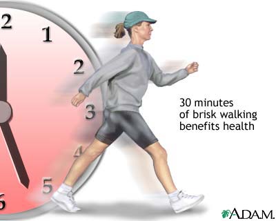
Aerobic exercise uses slow-twitch muscle fibers. Such exercise includes activities that are prolonged and require constant energy. Long distance running and cycling are examples of aerobic exercise. In aerobic exercise, the muscle cell requires the same amount of oxygen that the body supplies. The oxygen debt is slashed and lactic acid is not formed.
Skin
Introduction
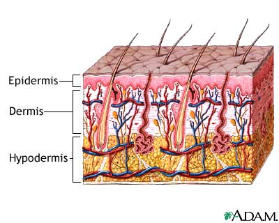
This most extensive organ system has the skin and accessory structures, including hair, nails, glands (sweat and sebaceous), and specialized nerve receptors for stimuli (changes in internal or external environment) such as touch, cold, heat, pain, and pressure. Its functions include protection of internal structures, prevention of entry of disease-causing microorganisms, temperature regulation, excretion through perspiration, pigmentary protection against ultraviolet sunrays, and production of vitamin D. The body stores about half its fat in the underlying hypodermis.
Skin: epidermal layers
The skin is the largest organ of the body, with a surface area of 18 square feet. Its two main layers are the epidermis (outer layer) and dermis (inner layer). The epidermis has several strata (layers) that contain four cell types. Keratinocytes produce keratin, a protein that gives skin its strength and flexibility and waterproofs the skin surface. Melanocytes produce melanin, the dark pigment that gives skin its color. Merkel's cells are probably involved with touch reception. Langerhans' cells help the immune system by processing antigens (foreign bodies).
The deepest layer of the epidermis, the stratum basale, is a single layer of cells resting on a basement membrane (layer between the dermis and epidermis). The stratum basale cells divide continuously. As new cells form, older ones are pushed toward the skin surface.
The epidermis does not have a direct blood supply; all nutrients that feed these cells come from the dermis. Only the deepest cells of the stratum basale receive nourishment. The cells that are pushed away from this layer die. When the cells reach the skin surface, they are sloughed off in a process called desquamation.
The next layer, the stratum spinosum, consists of spiny prickle cells that interlock to support the skin. The stratum granulosum, the thin middle layer, initiates keratinization (production of keratin). This process starts the death of epithelial cells (the cell type that makes up skin).
During desquamation, keratinocytes are pushed toward the surface. These cells begin to produce the keratin that eventually will dominate their contents. When these cells reach the epidermis outer layer, they are little more than keratin-filled sacs. Millions of these dead cells are worn off daily, creating a new epidermis every 35 to 45 days.
The stratum lucidum protects against sun ultraviolet-ray damage. This thick layer appears only in frequently used areas such as palms of the hands and soles of the feet. Thick skin epidermis has all five strata. Thin skin covers thinner epidermal areas such as eyelids. Thin skin has three or four of the five strata; it never has stratum lucidum.
The stratum corneum, the fifth, outermost layer is thick with rows of dead cells. These cells contain soft keratin, which keeps the skin elastic and protects underlying cells from drying out.
Skin: dermal layers
The dermis, called "true skin, " is the layer beneath the epidermis. Its major parts are collagen (a protein that adds strength), reticular fibers (thin protein fibers that add support), and elastic fibers (a protein that adds flexibility). The dermis has two layers: the papillary layer, which has loose connective tissue, and the reticular layer, which has dense connective tissue. These layers are so closely associated that they are difficult to differentiate.
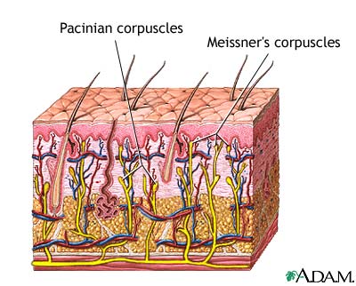
The papillary layer lies directly beneath the epidermis and connects to it via papillae (finger-like projections). Some papillae contain capillaries that nourish the epidermis; others contain Meissner's corpuscles, sensory touch receptors. A double row of papillae in finger pads produces the ridged fingerprints on fingertips. Similar patterns in the ridged fingerprints on fingertips are on palms of the hands and soles of the feet. Fingerprints and footprints keep skin from tearing and aid in gripping objects.
The reticular layer of the dermis contains criss-crossing collagen fibers that form a strong elastic network. This network forms a pattern called cleavage (Langer's) lines. Surgical incisions that are made parallel to cleavage lines heal faster and with less scarring than those made perpendicular. Parallel incisions disrupt collagen fibers less and require less scar tissue (cells that aid in healing) to close up a wound.
The reticular layer also contains Pacinian corpuscles, sensory receptors for deep pressure. This layer contains sweat glands, lymph vessels, smooth muscle, and hair follicles, described in the discussion on hair follicles later in this overview.
The hypodermis (subcutaneous layer) lies beneath the dermis. Loose connective tissue such as adipose tissue (fat) insulates the body, conserving heat. It also contains blood vessels, lymph vessels, and the bases of hair follicles and sweat glands. The fat distribution in this layer gives the female form its characteristic curves.
Sudoriferous (sweat) and sebaceous (oil) glands
Skin produces associated structures such as sudoriferous (sweat) glands and sebaceous (oil) glands. It also produces fingernails, hair, and sensory receptors that enable humans to feel pressure, temperature, and pain.
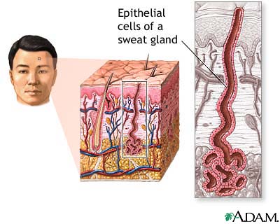
Both groups of sudoriferous glands (sweat glands) are in most of the body: eccrine glands are coiled ducts deep in the skin that connect to the surface; apocrine glands are in armpits, areolae of nipples, and the genital region. Eccrine glands secrete sweat, a mixture of 99 percent water and 1 percent salts and fats. In warm conditions with low humidity, perspiration (secretion of sweat) and evaporation cool the body.
Apocrine glands, which become active at puberty, are larger, deeper, and produce thicker secretions than eccrine glands. The apocrine glands secretions contain pheromones, substances that enable olfactory (sense of smell) communication with other members of the species. This communication provokes certain behavioral responses such as sexual arousal. Unlike eccrine glands that respond to heat, apocrine glands respond to stress and sexual activity by secreting sweat with a characteristic odor. This odor differs from body odor that results from bacteria decomposing skin secretions on the skin.
Ceruminous glands are modified apocrine glands in the external ear canal lining. They secrete cerumen (earwax), a sticky substance that is thought to repel foreign material.
Mammary glands in female breasts are modified apocrine glands. These glands are adapted to secrete milk instead of sweat.
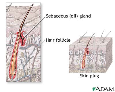
Sebaceous glands (oil glands) are all over the body except on the palms of hands and soles of feet. The glands empty via ducts into the bases of hair follicles and secrete sebum (a mixture of fats, waxes, and hydrocarbons). Sebum keeps hair moist and prevents skin from drying. Sebaceous glands are numerous on the face and scalp. During puberty, increased sex hormone levels in the blood may produce excessive sebum. This over secretion plugs the gland and hair follicle, producing a skin disorder called acne.
Hair and nails
Hair is composed of cornified threads of cells that develop from the epidermis and cover most of the body. Each hair has a medulla, cortex, and cuticle. The medulla in the center contains soft keratin and air. The cortex, the innermost thickest layer, has the pigment that gives hair color. The cuticle, the outermost layer, has cells that overlap like scales. Both the cuticle and cortex have hard keratin.
The hair root in a hair follicle is embedded beneath the skin. The hair shaft protrudes from the skin. Hair sheds and is replaced constantly during growth and rest phases. Hair has a protective function: eyebrows keep sweat from running into the eyes, nose and ear hairs filter dust from the air, and scalp hairs protect against abrasion and overexposure to sun rays.
Hair follicles extend into the dermis; the deep ends expanded parts are called hair bulbs. A papilla (connective tissue protrusion that contains capillaries) protrudes into the hair bulb and provides nutrients for the growing hair. The hair follicle walls have an inner epithelial root sheath and an outer dermal root sheath. The epithelial root sheath has an inner and an outer layer that thins as it approaches the hair bulb. It becomes the matrix, the actively growing part of the hair bulb that produces the hair.
Arrector pili muscles are smooth muscle cells attached to hair follicles. When they contract, they pull the hair into an upright position, causing skin dimples (goose bumps). The nervous system regulates these muscles; cold temperatures or fright can activate them.
Hair development begins in the third fetal month. By the fifth month, lanugo (thin hair) covers the fetus. At 5 months, lanugo disappears from every area except the scalp and eyebrows where coarser hair replaces it. Vellus (a film of delicate hair) eventually covers the rest of the body. Terminal hair is the early coarse scalp and eyebrow hair and later armpit and genital hair that grow during puberty. No new hair follicles develop after birth.
Like hair, nails develop from the epidermis. These hard plates of keratinized cells are at the ends of fingers and toes. Nails appear pink because their translucency reveals the vascular tissue beneath. They aid in grasping objects, scratching, and protecting fingers and toes.
The components of the nail are the lunula, body, root, and free edge. The lunula is the white half-moon shaped part at the nail base. Both the body and free edge region that overhangs the end of the finger or toe are visible. The nail rests on the thick layer of epithelial skin called the nail bed. The root is hidden under skin folds. Under the root lies the matrix (thick layer of skin). Eponychium (thin layer of epithelium) covers the nail during development; in the adult, it remains at the nail base only and is called the cuticle. The hyponychium is the epithelium of the nail bed.
Skin color
Skin color results from the presence of melanin, carotene (yellow to orange pigment), and underlying blood reflected through skin. Melanin keeps excessive ultraviolet rays from burning the skin. Exposure to sunlight causes the skin to produce more melanin, causing suntan, a temporary change in skin color. Melanin-rich cells continually move toward the surface, where they are sloughed. Too much sun is dangerous to skin; it increases the risk of cancer by affecting the genetic material of cells.
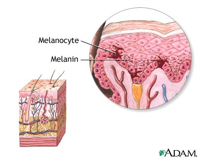
Variety of skin color is caused mainly by the number and distribution of melanocytes. Darker skin has more melanin that is produced by more melanocytes. However, the different skin colors among individuals and races do not reflect different numbers of melanocytes; instead, they show different kinds and amounts of melanin production by melanocytes. Oriental skin has a greater amount of carotene in the stratum corneum, producing a yellowish tinge. Albinism is a condition where skin does not produce melanin.